New Research : IIT Ropar studies about development of portable, low cost, an active infrared screening system, which will provide an early detection of breast cancer.
Navneet Kumar Gupta
Newswave @ Ropar
 IIT Ropar has introduced a novel pulse compression favourable Active Infrared Thermography for early detection of breast cancer in women of all ages, including pregnant or nursing women, irrespective of breast type (either fatty or dense breast).
The proposed technique makes use of infrared emission emanating from the breast to detect the hidden tumors inside it at very early stage for predefined thermal stimulus on to the breast under examination.
Infrared Thermography (IRT) is a fast, painless, non-contact, and non-invasive imaging method, complementary to mammography, ultrasound, and magnetic resonance imaging methods for early diagnosis of breast cancer. Widely used mammography shows its limitations in detecting tumours present in the dense breast.

Dense breasts have less fat and more gland tissue in comparison to the fatty breasts, which restricts mammography to detect tumours with confidence. Especially for the tumours situated in the gland region of breast due to the insignificant density variations between the gland and tumour regions, mammography fails to provide enough radiographic contrast between the tumour location and healthy region of the breast.
This limits the applicability of mammography in screening of dense breasts. Also, mammography provides discomfort to the patient and the exposure to harmful ionizing radiation further restricts its applicability. However, the present Active IRT technique outperforms the standard method of mammography by providing patient friendly breast screening.
The present study carried out at IIT Ropar emphasis on Active InfraRed Thermography/Thermal Wave Imaging technique to detect tumors with significant thermal contrast over the breast. In this approach, an external thermal stimulus (heat/cold) is applied over the breast under examination for creating significant temperature differences (2 to 3 degrees centigrade from the ambient breast temperature) over the normal and abnormal (tumour) regions over the breast.
The thermal stimulus can be applied either in a pulsed form (pulse and pulse-phase thermography) or in a harmonic modulated way (lock-in thermography). Pulse thermography (PT) demands a higher peak power source and has the additional drawback of non-uniform heating. In lock-in thermography (LT), to detect the abnormalities present at different depths of various spatial dimensions in the breast under examination, demands the repetition of the experiment at different excitation frequencies to resolve them with enough thermal contrast, which makes this technique a time consuming process.
The present research focuses on pulse compression favourable linear frequency modulated thermal wave imaging (LFMTWI) developed by Mulaveesala et al. a decade back for testing and evaluation of various industrial materials. Using LFMTWI technique, a frequency-modulated heat stimulus with frequencies varying within a predefined band having equal magnitude is imposed over the breast.
Thermal waves generated due to applied heat stimulus diffuse into the breast and produce a similar temporal temperature distribution on the skin surface of the breast with a certain delay and amplitude. The presence of tumors inside the breast alters the heat flow resulting in temperature gradients over the surface. Further, phase and amplitude images are constructed using frequency and time-domain data analysis schemes for detecting the sub-surface tumours with improved contrast.
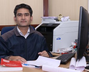
Dr. Ravibabu Mulaveesala, Associate Professor, Deptt of Elect Engg, IIT Ropar, said, “Following success of research predictions of our group at InfraRed Imaging Laboratory (IRIL), we are now working towards the development of portable, low cost, an active infrared screening system, which will provide an early detection of breast cancer irrespective of patient’s age, size, type of breast (either fatty or dense) and it’s stage”.
The team of researchers is led by Dr. Ravibabu Mulaveesala, Associate Professor, InfraRed Imaging Laboratory (IRIL), Deptt of Elect.Engg, IIT Ropar. The research paper, ‘Applicability of active infrared thermography for screening of human breast: a numerical study’, has been published in Journal of Biomedical Optics.
 News Wave Waves of News


